
Figure 4.
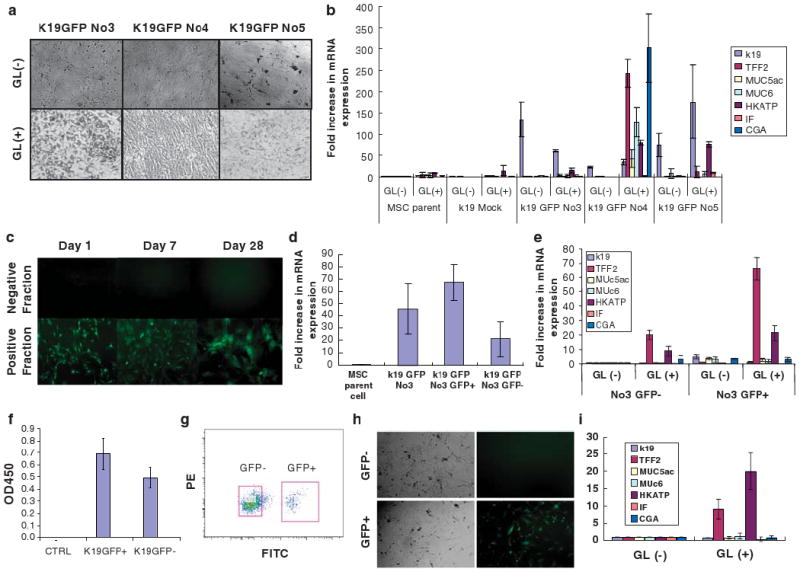
Induced expression of gastric phenotype markers in K19-positive MSC clones after treatment with gastric tissue extract. (a) K19GFP MSC clone no. 3, no. 4, and no. 5 were incubated with gastric tissue extract (GL) for 5 days. The morphology of K19GFP MSC clones no. 3, no. 4, and no. 5 after treatment is shown. Original magnification, × 100. (b) Expression of gastric epithelial phenotype markers, such as k19, TFF2, Muc5ac, Muc6, H/K-ATPase, IF, and CGA in K19GFP MSC clones after treatment with GL assessed by real-time PCR. Average data of three separate experiments is shown. (c) Sorted GFP+ and GFP− cells of K19GFPMSCNo3 were cultured separately, and the expression of GFP was assessed after 1, 7, and 28 days of culture. GFP− cells could not generate GFP+ cells. Original magnification, × 100. (d) Fold increase in K19 mRNA expression level in pooled K19GFP No3, sorted GFP+ and GFP− fraction were compared with that of parent cells by real-time PCR. (n = 3). (e) The expression of gastric epithelial phenotypic markers, such as K19, TFF2, Muc5ac, Muc6, H/K-ATPase, IF, and CGA in the sorted GFP+ and GFP− cells after treatment with GL were assessed by real-time PCR. (f) BrdU assay using K19GFP MSC clone no. 3 revealed a higher proliferation ability of GFP+ MSCs over GFP− MSCs (eight samples each, unpaired Student's t-test P = 0.0029). (g) GFP+ and GFP− cells were isolated by fluorescent cell sorting from K19GFP no. 3. (h) Colony-forming ability of single-sorted GFP+ and GFP− cells isolated from K19GFP MSC clone no. 3. GFP+ cells could give rise to both GFP+ and GFP− cells, whereas GFP− cells could not generate GFP+ cells. Original magnification, × 100. (i) The expression of gastric epithelial phenotypic markers, such as K19, TFF2, Muc5ac, Muc6, H/K-ATPase, IF, and CGA in the colonies derived from single-sorted GFP+ and GFP− cells after treatment with GL were assessed by RT-PCR.