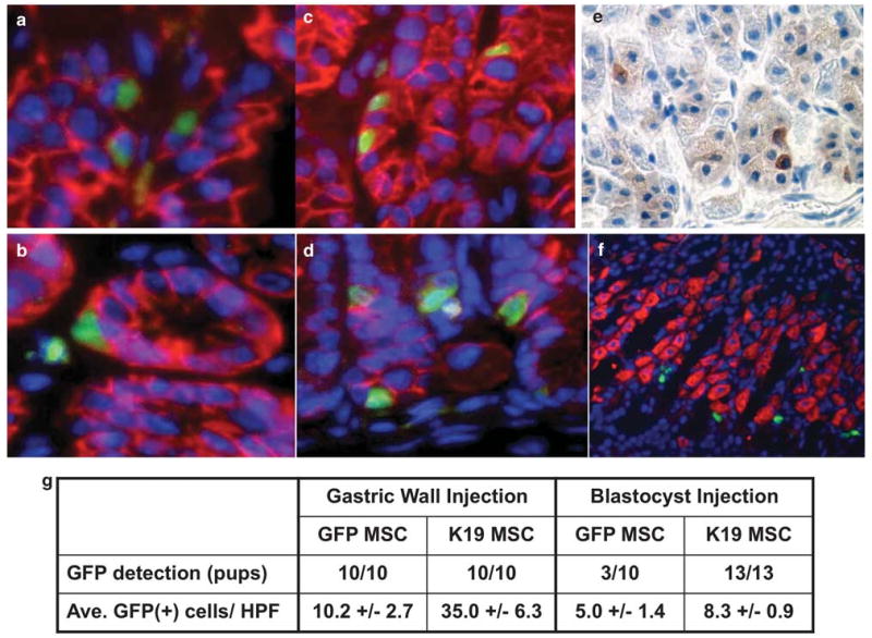
Figure 5.

Contribution of K19-positive MSCs to the gastric epithelium in vivo. (a) GFPMSCs (200 000 cells in 10 μl of PBS) were injected into the gastric wall of C57BL/6 mice and gastric tissue sections were prepared 2 weeks after injection. Sections were stained with an anti-E-cadherin antibody and Texas Red-conjugated secondary antibody. Nuclei were stained with DAPI. Original magnification, × 400. (b) K19GFP MSC no. 4 (200 000 cells in 10 μl of PBS) were injected into the gastric wall of C57BL/6 mice and gastric tissue sections were prepared 2 weeks after injection. Sections were stained with an anti-E-cadherin antibody and Texas Red-conjugated secondary antibody. Original magnification, × 400. (c) GFPMSCs were injected into 3.5 day-old mouse blastocysts to establish chimeric mice and gastric tissue sections were prepared at 8 weeks of age. Sections were stained with an anti-E-cadherin antibody in combination with Texas Red-conjugated secondary antibody. Nuclei were stained with DAPI. Original magnification, × 400. (d) K19GFP MSC no. 4 was injected into 3.5 day-old mouse blastocysts to establish chimeric mice and gastric tissue sections were prepared at 8 weeks of age. Sections were stained with an anti-E-cadherin antibody and Texas Red-conjugated secondary antibody. Nuclei were stained with DAPI. Original magnification, × 400. (e) Immunohistochemistry with an anti-GFP antibody. Representative stomach section of a mouse derived from blastocyst injection of GFPMSC is shown. Original magnification, × 300. (f) Immunofluorescence of H/K-ATPase and GFP-positive cells. Sections were stained with an anti H/K-ATPase antibody and Texas Red-conjugated secondary antibody. Nuclei were stained with DAPI. A representative stomach section of a mouse derived from blastocyst injection of GFPMSC is shown. Original magnification, × 200. (g) GFP+ cell detection rate and GFP+ cells per high-power field (HPF) in both gastric wall injection and blastocyst injection studies.
