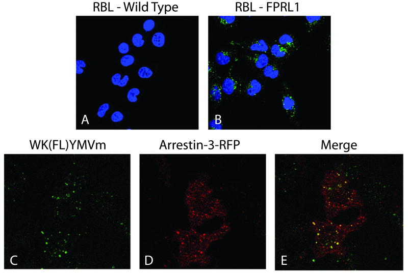
Figure 3.
Internalization of fluorescent ligand bound FPRL1 and colocalization with arrestin-3 in RBL cells. The cells were all exposed to 5 nm WK(FL)YMVm for 10 min at 37°C before being washed and fixed. A,B: Internalization of the fluorescent WK(FL)YMVm ligand (green) is demonstrated in wild-type RBL cells (A) and FPRL1 expressing cells (B). Nuclei are stained with DAPI (blue). C_E: Colocalization of fluorescent ligand with arrestin-3. RBL cells transiently transfected with RFP-arrestin-3 (D) were allowed to internalize ligand as before (C); merged image in E.

