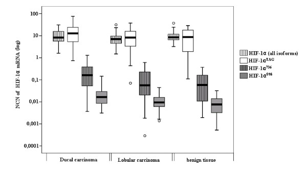
Figure 2.
Distribution of hypoxia inducible factor 1α (HIF-1α) mRNA levels in 82 breast tissues determined by real-time quantitative reverse transcription PCR assay. HIF-1α mRNA levels were expressed in normalised copy numbers (NCN) on the basis of TATA box-binding protein (TPB) gene content of the tissues as described in the Methods section. Patients grouped according to histological type of tissues (x axis). HIF-1α mRNA levels (y axis) were represented according to these different groups of patients. Results were plotted on a logarithmic scale. HIF-1α splice variants were detectable in all samples at varying levels. Splice variant HIF-1α736 and HIF-1α516 mRNAs were expressed at levels 100-fold lower than HIF-1αTAG. Variant HIF-1α557 mRNAs that were expressed at levels 1,000-fold lower than HIF-1αTAG are not shown.

