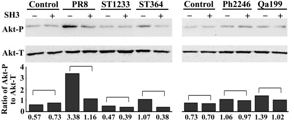
Figure 3.
Western blot analysis of phosphorylated Akt level in MDCK cells. MDCK cells with or without heterologous SH3 were infected with PR8, ST1233, ST364, Ph2246, or Qa199 at an MOI of 2 for 8 h. Akt-P (phosphorylated Akt at Serine 473) and Akt-T (total Akt) in cellular lysate were detected using anti-Akt-P and anti-Akt-T antibodies, respectively, and peroxidase-conjugated secondary antibody.

