
Figure 2.
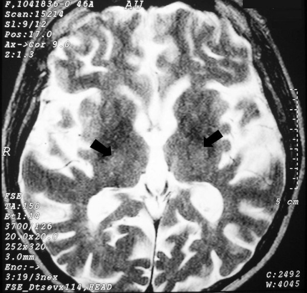
Brain axial T2W magnetic resonance imaging at the level of the thalami and third ventricle. High signal foci in the medial regions of thalami (arrows).
Official websites use .gov
A
.gov website belongs to an official
government organization in the United States.
Secure .gov websites use HTTPS
A lock (
) or https:// means you've safely
connected to the .gov website. Share sensitive
information only on official, secure websites.
Brain axial T2W magnetic resonance imaging at the level of the thalami and third ventricle. High signal foci in the medial regions of thalami (arrows).