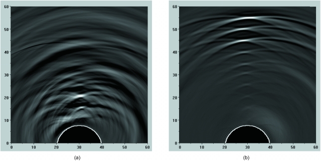
Figure 2.
Images of combined OA signals for 1-mm cylindrical tube (with the tube axis oriented orthogonal to the imaging plane) positioned at various depths within the aqueous medium simulating optical properties of background tissues, and illuminated using (a) backward OA mode and (b) forward OA mode. OA signals were acquired using a single target, and the images were subsequently created after combining the signals from each individual acquisition. The images are displayed using the standard 8-bit gray-scale palette. The axes are scaled in millimeters.

