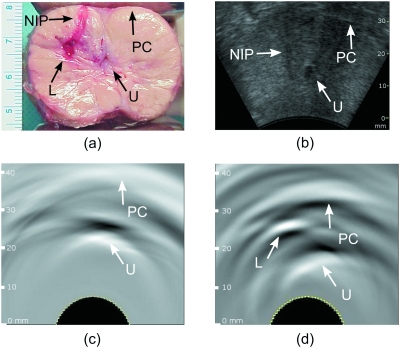
Figure 4.
(a) Photograph of the sliced dog prostate showing the presence of the induced lesion with blood in the right lateral lobe, the urethra is visible but contracted after surgical excision; (b) ultrasonic image of the same dog prostate obtained in vivo after the surgery. OA images of the same dog prostate obtained in vivo (c) before and (d) after the lesion was induced. The induced bloody lesion can be seen in (a) and (d). The needle insertion path is visible in (a) and (b). Due to the acoustic mismatch between tissue and air, the prostate capsule can be identified in OA images as a white band. Arrows indicate the prostate capsule (PC), urethra (U), needle insertion path (NIP), and lesion (L). The images are displayed using the standard 8-bit gray-scale palette.

