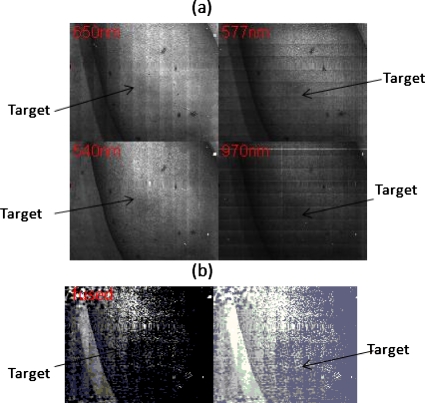
Figure 3.
Real-time acquired images of an erythema on a dark pigmented arm. The four images correspond to four different wavelengths λ[1,2,3,4]. (b) Left side shows the fused view of the erythema. The fusion algorithm is given by Eq. 1. Right shows the same as the left, but with a histogram equalization procedure being applied.

