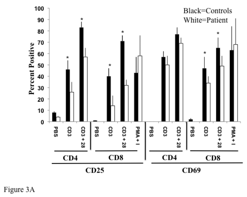
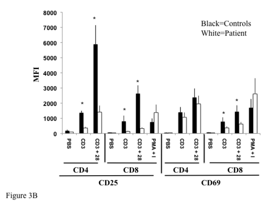
Fig 3. Reduced expression of activation markers by T cells from IRAK-4 deficient patients.
Control and patient non-adherent lymphocytes were cultured with PBS (Ø), immobilized anti-CD3 (CD3), immobilized anti-CD3 plus soluble anti-CD28 (CD3/28), or PMA and ionomycin and stained as in Fig 2. Data from four patients and controls was averaged and plotted as percent positive (A), and mean fluorescence intensity (MFI) (B). *p < 0.05 by student t test.


