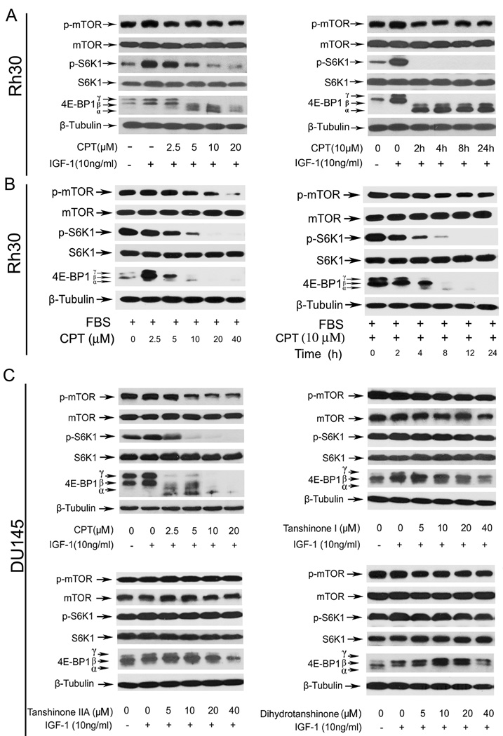
Fig. 4. CPT inhibits mTOR pathway in cancer cells.
Western blot analysis was performed for (A–D). β-Tubulin was used for loading control. Serum-starved (A) or non-starved Rh30 cells (B) were pre-treated with CPT (0–40 µM) for 2 h, and then stimulated with IGF-1 (10 ng/ml) for 1 h (Left Panel), or with CPT (10 µM) for the indicated time and stimulated with IGF-1 (10 ng/ml) for 1 h (Right Panel). (C) Serum-starved DU145 cells were treated with CPT, tanshinone I, tanshinone IIA or dihydrotanshinone (0–40 µM), respectively, for 2 h, and then stimulated with IGF-1 (10 ng/ml) for 1 h.

