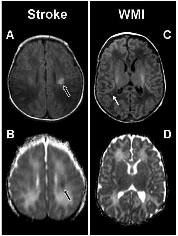
Figure 1. Patterns of Brain Injury: stroke and white matter injury (WMI).

The abnormal hypersignal (black arrow) in the left basal ganglia is an example of a small preoperative stroke in a term newborn with TGA. It is localized in the middle cerebral territory and appears as a hyperintensity on the (A) T1-weighted image and an area of restricted diffusion on the (B) ADC map. The images on the right are an example of preoperative white matter injury (WMI) in a term newborn with transposition of the great arteries (TGA), and show a small focus (white arrow) of hyperintensity in the right parietal lobe on (C) the axial T1-weighted imaging. There is no corresponding area of restricted diffusion on (D) the ADC map.
