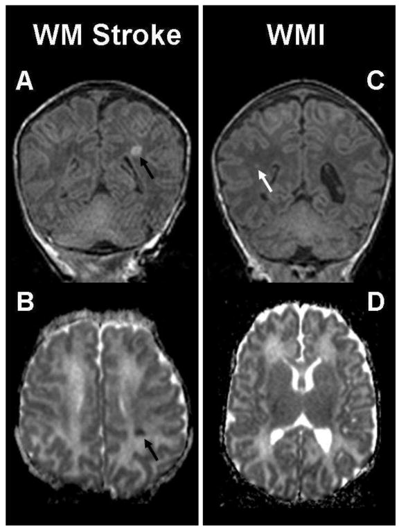
Figure 2. Solitary white matter lesions.

The coronal T1-weighted imaging (A) and axial ADC map (B) show a small preoperative white matter stroke (black arrow) in a term newborn with transposition of the great arteries (TGA). The lesion is localized in the left middle cerebral artery territory and characterized by an abnormal T1 hyperintensity (A) and an area of restricted diffusion (B) in left parietal lobe. The images on the right are an example of white matter injury (WMI) in another term newborn with TGA (same patient as in Figure 1), and show a small focus (white arrow) of hyperintensity in the right parietal lobe on (C) the coronal T1-weighted imaging. There is no corresponding area of restricted diffusion on (D) the ADC map.
