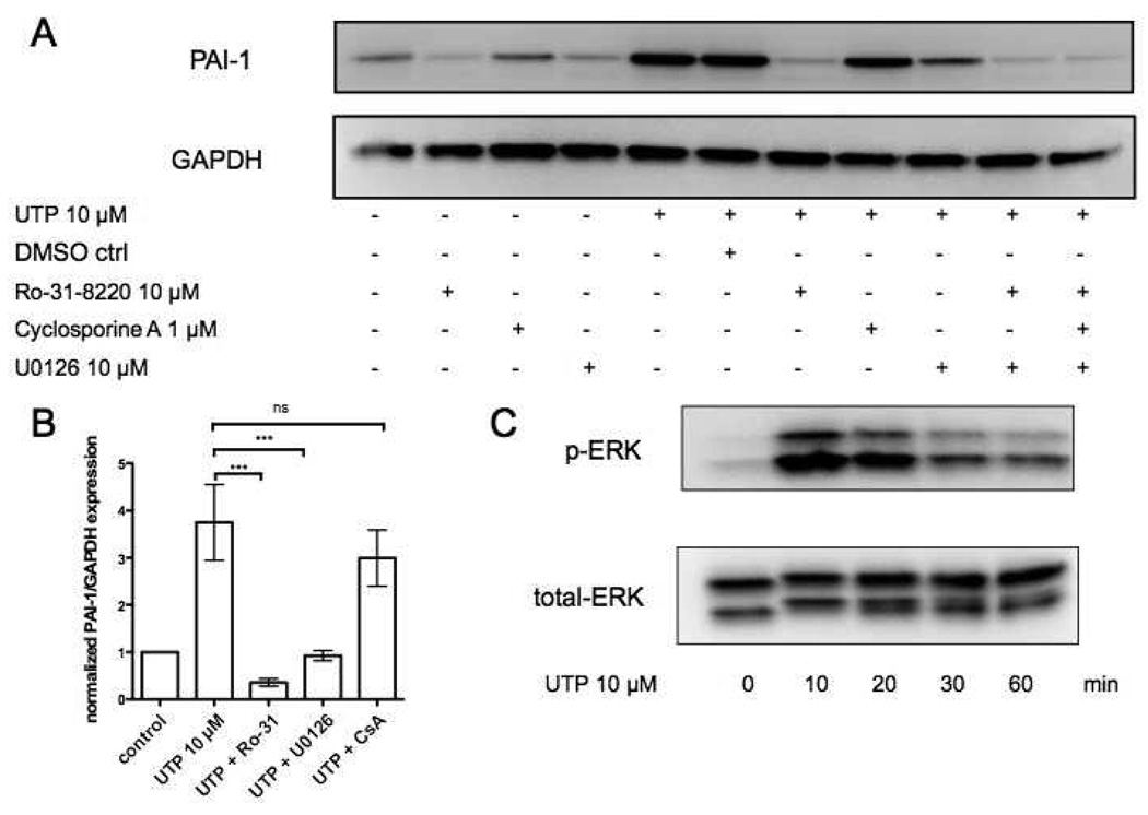
Figure 5. UTP induces PAI-1 expression in rat cardiac fibroblasts in a PKC and ERK-dependent manner.
(A) CFs were serum-starved for 24h and then incubated in the presence or absence of: DMSO (vehicle control), Ro-31-8220 (PKC inhibitor), U0126 (MAPK/ERK kinase (MEK) inhibitor), or Cyclosporine A (calcineurin inhibitor) for 30 min. The cells were then stimulated with UTP (10µM, 4 h). Cells were lysed and assayed for PAI-1 protein expression. GAPDH was used to normalize for protein loading. Panel (B) shows quantification of the immunoblots from panel (A). (C) ERK-phosphorylation and total ERK protein were assessed using immunoblots following stimulation with 10 µM UTP for 0, 10, 20, 30, 60 min. The data shown are mean ± SEM of at least 3 independent experiments performed in triplicate and compared by using ANOVA with post-hoc multiple comparison tests. ***, p<0.001.

