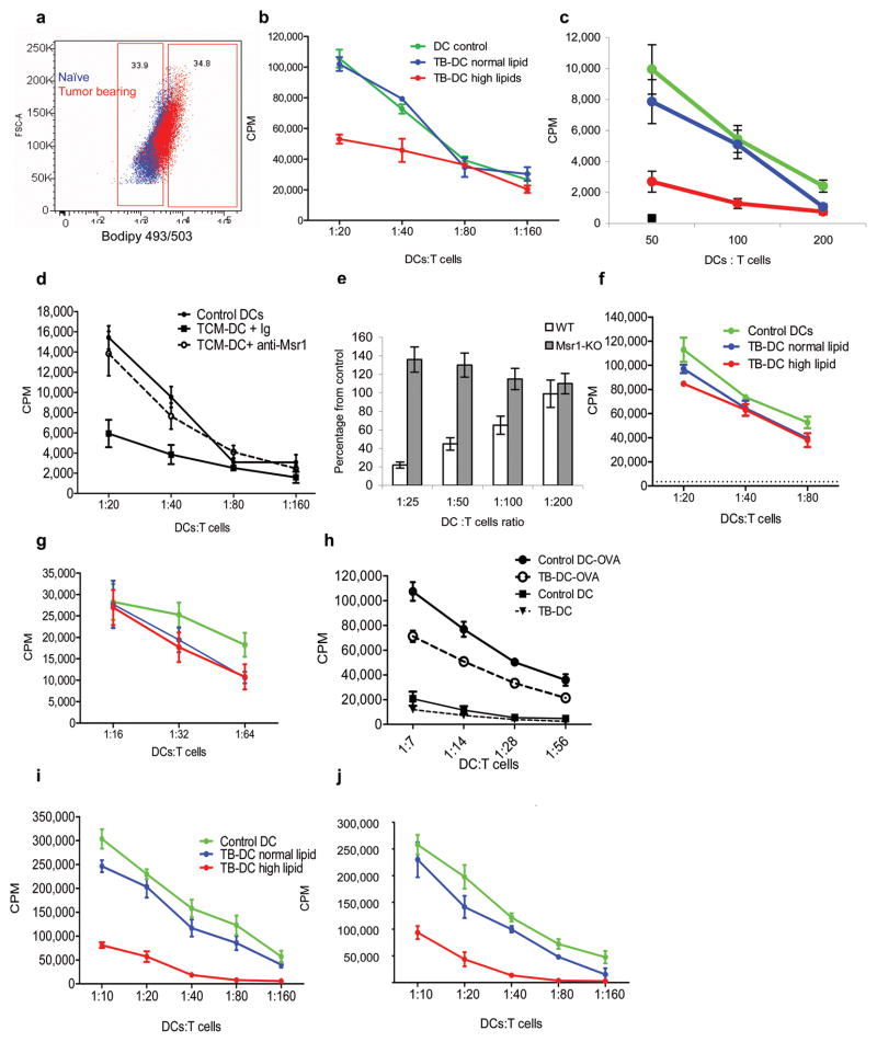
Figure 4. Defective functional activity of DCs with high lipid content.
a,b. Stimulation of allogeneic T cell proliferation by DCs with different lipid content isolated from CT-26 TB mice. a. Examples of gates set for the sort of CD11c+ cells. Blue- naïve mouse, red–TB mouse. b. Stimulation of proliferation of allogeneic T cells by sorted DCs with high and low lipid content. Typical results of 4 performed experiments are shown. T cells alone had 3[H]-thymidine uptake of less than 500 CPM. c. Stimulation of allogeneic T cell proliferation by DCs generated from enriched BM HPC from naïve BALB/c mice, exposed for 2 days to CT-26 TES and sorted for DC-HL (red) or DC-NL (green) and compared to DCs cultured in control medium (blue). Three experiments with similar results were performed. T cells alone had 3[H]-thymidine uptake of less than 200 CPM. d. DCs were generated from HPC of C57BL/6 mice and cultured for 3 days with EL-4 TES in the presence of control IgG or anti-MSR1 antibody. After that time cells were cultured in triplicate with allogeneic T cells at indicated ratios and cell proliferation was evaluated by 3[H]-thymidine uptake. T cells alone had 3[H]-thymidine uptake of less than 500 CPM. e. DCs generated from BM HPC of wild-type and MSR1 KO mice in the presence of TES as described in Fig. 3j were used as stimulators in an allogeneic mixed leukocyte reaction. T-cell proliferation was measured by 3[H]-thymidine uptake. Two independent experiments were performed in triplicate. The values of T-cell proliferation induced by wild-type or MSR1-deifient DCs generated without the presence of TES were set as 100%. Values of T-cell proliferation induced by DCs generated in the presence of TES are presented as percentage from control level. f,g. DCs with high and normal lipid content were sorted from spleens of naïve and EL-4 TB mice and incubated at indicated ratios with T cells from OT-II (f) or OT-I (g) transgenic mice in the presence of 10 μg/ml of specific peptides. T cell proliferation was measured in triplicate by 3[H]-thymidine uptake. In the absence of peptides 3[H]-thymidine uptake in all samples was less than 5×10³ CPM. Three experiments with the same results were performed. h. CD11c+ DCs isolated from spleens of naïve and EL-4 TB mice were cultured overnight with 1 mg/ml OVA or in medium alone and then used to stimulate T cells from OT-II transgenic mice. T cell proliferation was measured in triplicate by 3[H]-thymidine uptake. i.j. DCs with high and normal lipid content were sorted from the spleens of EL-4 TB mice and incubated overnight with 1 mg/ml OVA. Cells were used to stimulate T cells from OT-II (i) and OT-I (j) transgenic mice. T-cell proliferation was measured in triplicate using 3[H]-thymidine uptake. Two experiments with similar results were performed.

