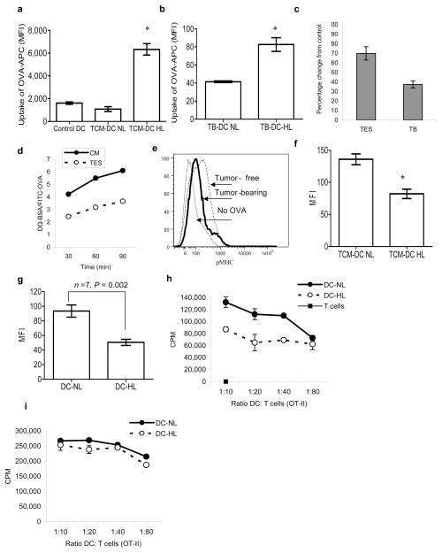
Figure 5. Antigen processing in lipid-laden DCs.
a. One-hour uptake of OVA-APC by DCs generated from HPC in the presence of TES. The level of APC fluorescence was evaluated within the DC-NL and DC-HL populations. Mean±SD of three performed experiments are shown. * - statistically significant differences (p<0.05) from control DCs. b. Uptake of OVA-APC by DCs from EL-4 TB mice 2 h after i.p. injection of 100 μg of OVA-APC. Mean±SD of three mice per group is shown. c. DCs were either generated in vitro and then cultured for 48 h in control medium or TES, or isolated directly from spleens of naïve or EL-4 tumor-bearing (TB) mice. Cells were loaded with either 1 mg/ml DQ-BSA or 10 mg/ml FITC-OVA. DQ-BSA/FITC-OVA ratio was calculated within CD11c+ DCs after 1 h incubation. DQ-BSA/FITC-OVA ratio in control samples was expressed as 100%. Two experiments with the same results were performed. d. DCs generated in vitro as described above were loaded with DQ-BSA or FITC-OVA and incubated for the indicated time. Fluorescence in CD11c+ DCs was evaluated as described above. e. CD11c+ DCs were isolated from spleens of tumor-free and EL-4 tumor-bearing mice and loaded overnight with 1 mg/ml OVA. Cells were then stained with APC conjugated 25-d1.16 antibody. A typical result of one experiment out of 4 performed is shown. f. DCs were generated from progenitors in the presence of EL-4 TES and then loaded overnight with 1 mg/ml OVA. DCs were stained with Bodipy 493/503 and labeled with 25-d1.16 antibody. DCs with high and normal lipid content were analyzed. The mean fluorescence of cells stained with isotype control Ig was less than 20. * - statistically significant differences (p<0.05) between DC-NL and DC-HL. Cumulative results of 3 independent experiments are shown. g. Binding of 25-d1.16 antibody to DC-NL and DC-HL DCs from LN cells of EG-7 TB mice. The fluorescence of cells stained with isotype control Ig was less than 20. h.i. DCs with high and normal lipid content were sorted from LN of EG-7 TB mice and incubated with T cells isolated from OT-II transgenic mice in the absence of (h) or in the presence of 10 μg/ml of OT-II specific peptide (i). T cell proliferation was measured in triplicate by 3[H]-thymidine uptake. Two experiments with the same results were performed.

