
Fig. 1. Dissolving polymer microneedle patches.
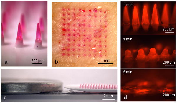
(a) Side view of dissolving polymer microneedles. (b) En face view of porcine skin after insertion and removal of microneedles, showing delivery of the encapsulated compound (sulforhodamine). (c) Relative height of microneedles next to a U.S. nickel coin. (d) Polymer microneedle dissolution in pig skin in vitro. Frame 1 = pre-insertion, frame 2 = after 1 min insertion in skin, frame 3 = after 5 min insertion in skin.