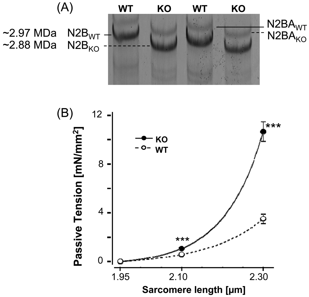
Figure 1. Characterization of the N2B KO mouse model.
(A) Titin expression in LV myocardium of WT and N2B KO mice (1% agarose gels). WT myocardium of the mouse expresses predominately N2B titin with a small level of N2BA titin. In the KO both N2B titin and N2BA titin have a slightly higher mobility than in the WT, consistent with the excision of the N2B element. (B) Titin-based passive tension in WT and N2B KO skinned myocardium (results from 8 WT and 8 KO mice). (Tension is steady-state tension and was measured after 5 min stress relaxation.) Asterisks: comparison between KO and WT myocardium.

