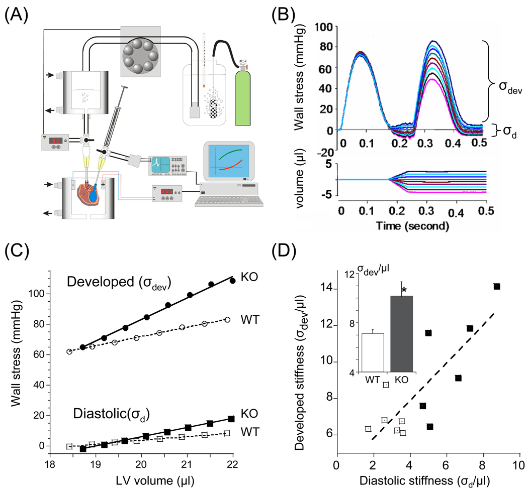
Figure 7. Assessment of FSM in isolated hearts.
(A) Left: Schematic of isolated heart setup. The heart is perfused, twitch activated, and a small fluid-filled balloon, introduced into the LV and connected to a fast servomotor, rapidly changes LV volume; a pressure sensor inserted in the balloon measure LV pressure. (B) Superimposed family of set of contractions before and after (test contraction) volume change. Pressure was converted to wall stress (σ) and developed stress (σdev) and diastolic stress (σd) were determined. (C) Example of results of control (open symbols) and KO heart (closed symbols) obtained in the presence of dobutamine. Note that the developed stress (σdev) – volume relation is steeper in the KO than in the WT heart. (D) Developed stiffness (slope of σ - volume relation) is positively correlated with diastolic stiffness (p<0.01). Individual results from WT and KO hearts are shown. Inset: comparison of mean values (asterisks: comparison between WT and KO). Results from 6 WT and 6 KO mice

