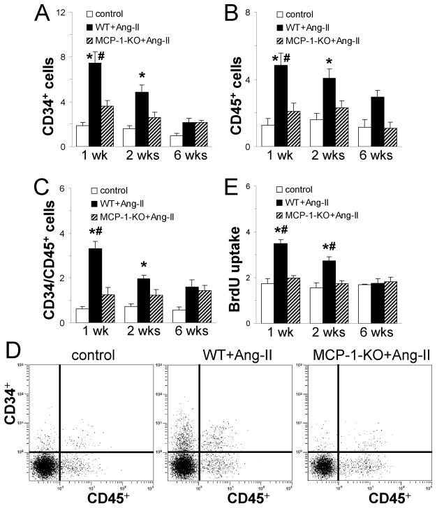
Fig. 3. Continuous Ang-II infusion induces the appearance of primitive, hematopoietic cells in the heart that is dependent on MCP-1 expression.
Hearts were removed and non-myocytes isolated. (A-C) Dispersed cells were analyzed for (A) CD34, (B) CD45, and (C) both CD34 and CD45 expression on viable (calcein+) cells by flow cytometry. (D) Representative cytometric diagrams showing the CD34+ and CD45+ distribution of all viable non-myocytes. Ang-II infusion results in the uptake of hematopoietic cells in WT mouse hearts. Deletion of MCP-1 reduces the number of CD34 and CD45 positive cells compared with WT. (E) The same cells were cultured in vitro. Ang-II infusion in WT mice results in increased cell proliferation (BrdU uptake) compared to control cultures. Cells isolated from Ang-II-treated MCP-1-KO mice proliferate at similar rates as isolations from control groups. Control mice received saline (1 and 2 wks: n=6 WT + n=2 MCP-1-KO; 6 wks: n=4 WT + n=1 MCP-1-KO; no difference between groups was observed); WT and MCP-1-KO mice (1 and 2 wks: n= 8/group; 6 wks: n=5/group) received Ang-II for the indicated time. *P<0.05 between Ang-II-treated and control groups; #P<0.05 between Ang-II-treated WT and MCP-1-KO groups.

