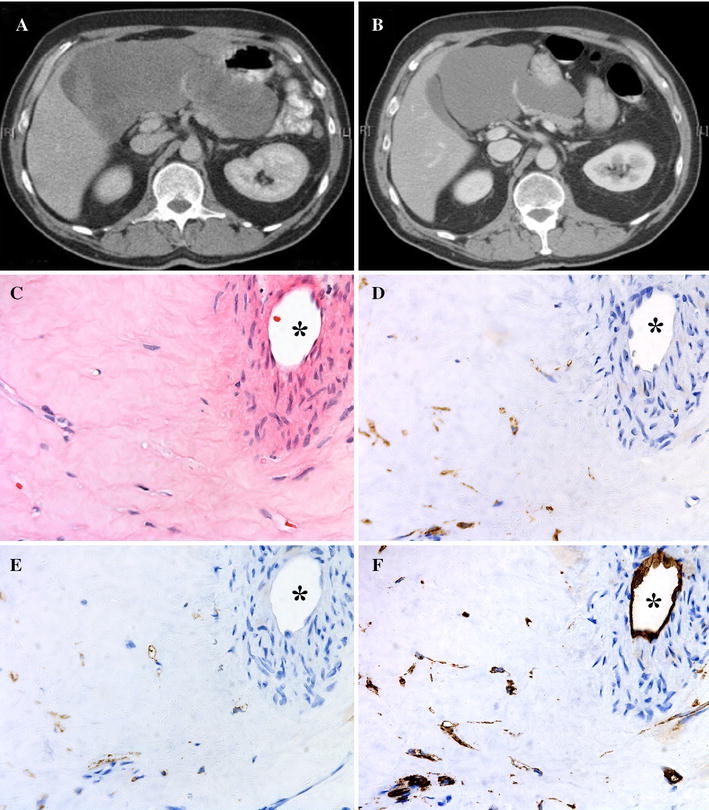
Fig. 2.

Computed tomography scan before and after 9 months of downsizing sunitinib. The 21 cm tumor (a) was reduced to 14 cm (b) in diameter prior to resection. c–f Histopathology of this tumor after sunitinib treatment. The tumor was composed mainly of collagen fibrous tissue with scattered tumor cells (c) and was positive for KIT (d), DOG1 (e), and CD34 (f). A major vessel is seen in the upper right corner (*)
