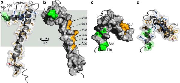
Fig. 3.
Molecular backbone and surface representations of FXYD1 (PDB accession code: 2JO1). In the helical regions, basic side-chains are shown in blue, acidic side-chains are red, and apolar side-chains are yellow. The three Ser residues (S62, S63, S68) and Thr69 in the cytoplasmic helix are in green. Residues in the transmembrane helix (G20, A24, G25, F28, G31, V35), predicted to interact with the Na,K-ATPase α subunit are shown in yellow. Side views. The structures are viewed (a, d) from the membrane side, or (c, d) down the membrane surface from the cytoplasm

