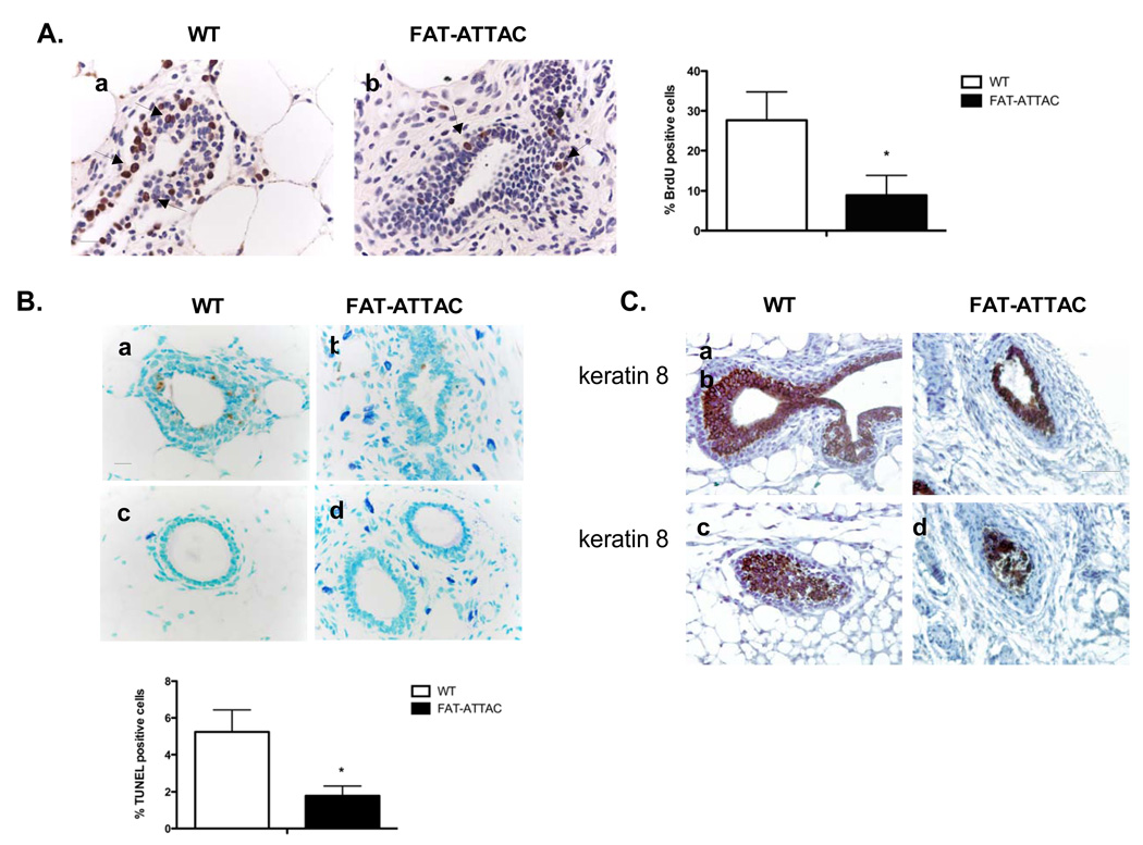
Figure 6. Ablation of mammary stromal adipocytes results in inhibition of both proliferation and apoptosis.
A) BrdU immunohistochemistry was carried out on mammary glands derived from mice that were treated with the dimerizer for a 2-week period. Shown is the % BrdU positive cells per TEB field. (*p<0.05, n=4/per group, Scale bar 20mm). B) TUNEL immunohistochemistry of mice dimerized for 4 weeks. Shown is quantification of the TUNEL immunohistochemistry per TEB field (*p<0.05, n=4/per group, Scale bar 20mm). C) Mice were dimerized for 4 weeks and immunohistochemical analysis of TEBs was conducted using the luminal epithelial marker cytokeratin 8 (n=5/per group, Scale bar 50mm).

