Figure 1.

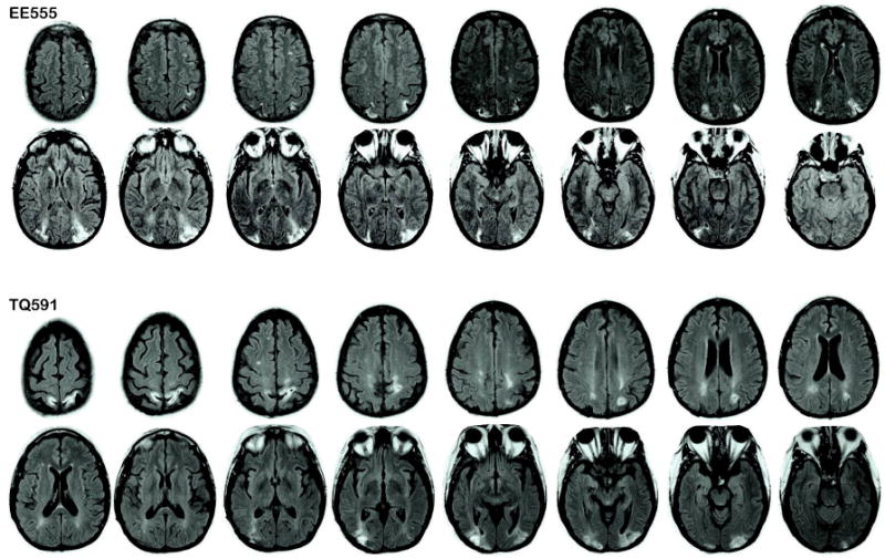
Patient lesion traces. Images are T2 fluid-attenuated inversion recovery (FLAIR) images in which the lesions appear as white higher intensity patches in parietal regions. Right is on the left.

Patient lesion traces. Images are T2 fluid-attenuated inversion recovery (FLAIR) images in which the lesions appear as white higher intensity patches in parietal regions. Right is on the left.