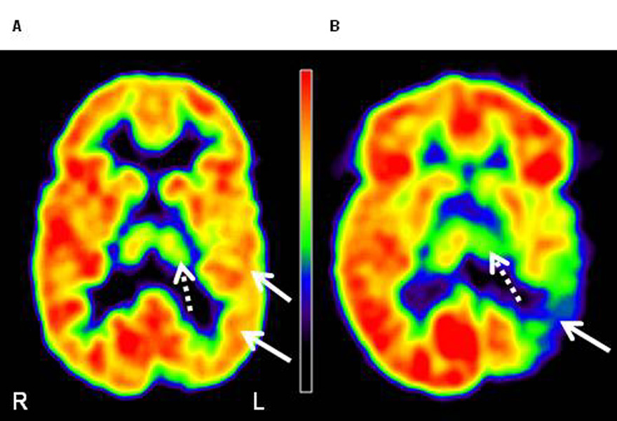
Figure 2.
FDG PET images of two patients with Sturge-Weber syndrome, with mild (A) and severe (B) left posterior (parietal, occipital, temporal) cortical involvement (solid arrows). (A) The image of a 4 years old boy (patient 4) shows minimal thalamic hypometabolism (dashed arrows; measured AI: −5.2%) on the left side. This patient had normal IQ (full-scale IQ: 110). (B) The image of an 8 years old girl (patient 5) shows severe thalamic hypometabolism (dashed arrow; AI: −23.7%). This patient`s cognitive functions were severely impaired (full-scale IQ: 55). L=left, R=right.

