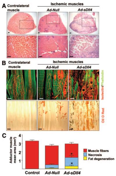
Figure 4. Dll4 Inhibition impairs muscle regeneration.
A, Cross-section showing the whole perimeter of adductor muscles harvested at 14 days postischemia and stained with hematoxylin/eosin. Lower panel: higher magnification of upper panel. B) Considerable loss of muscle fibers in Ad-sDll4–injected ischemic muscles. Myocytes (phalloidin, green), microvessels (IsolectinB4, red). Scale bar: 100 μm. Bottom images, Lipid deposit is increased in Ad-sDll4–injected ischemic muscles as revealed by oil red O staining (red). Scale bar: 200 μm. C, Quantification of muscle fibers, lipid degeneration, and necrosis area. *P<0.05; n=6 per group.

