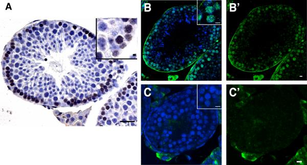
Fig. 4. Spo11 dependent p53 activation is conserved in the mouse.
(A–C) Immunostainings for phospho-Ser15 p53 in paraffin sections from wild-type (A, B) and Spo11 knockout (C) mouse testes. DAB chromogen (A, brown) and fluorescent (B,C, green) detection methods show transient activation of p53 in meiotic cells is absent in Spo11-deficient testes. DNA staining (hematoxylin or Hoechst) is in blue. B' and C' show signal without Hoechst counterstain. Insets in (B) and (C) show nuclear localized staining of Ser15-p53 under comparable magnifications. Scale bar, 100 μm (A); 10μm (B–C, insets in A).

