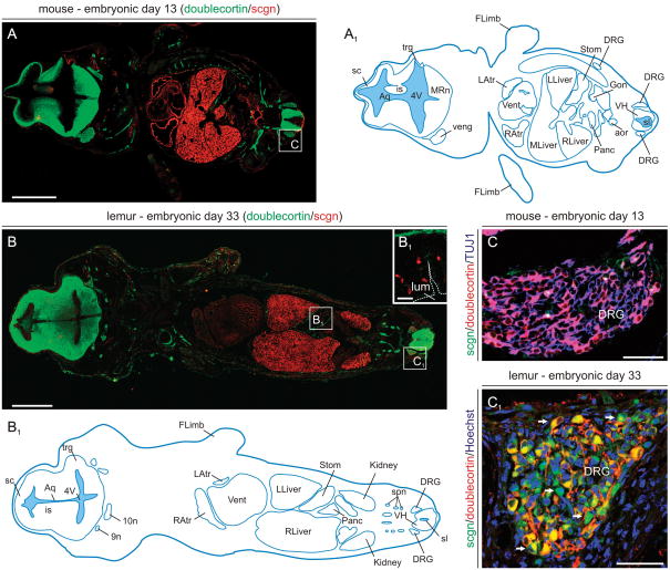
Fig. 2. Scgn expression in mouse and primate embryos at mid-gestation.
(A,A1) Scgn+ organ systems in mouse embryo. Doublecortin, a microtubule-associated protein widely expressed by migrating cells in the developing nervous system (Gleeson et al., 1999), was used to reveal anatomical boundaries of various organs. (B,B1) Scgn+ organs in the grey mouse lemur embryo. Open rectangles in A and B denote the general location of high-resolution photomicrographs in B1-C1. (B1) Scgn immunoreactivity decorates putative enteroendocrine cells (Mulder et al., 2009b) at the base of gastric pits formed in the stomach of lemur embryos. (C,C1) Prospective sensory neurons in the dorsal root ganglion (DRG) of the lemur (arrows, C1) but not mouse (C) express scgn. Abbreviations are listed in Supporting Table 1. Scale bars = 30 μm (B1), 150 μm (C,C1), 0.5 mm (A,B).

