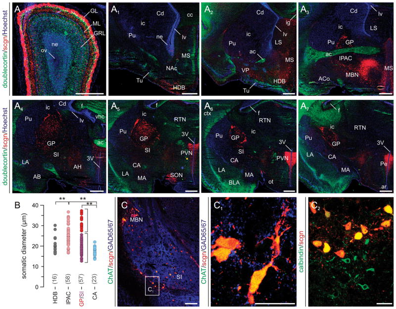
Fig. 6. Scgn distribution in fetal mouse lemur forebrain.
(A–A7) Scgn expression was identified in coronal sections with a sampling interval of 700 μm along the anterior-posterior axis of the fetal mouse lemur brain at day 50 of gestation. Doublecortin immunoreactivity together with nuclear counterstaining was used to identify anatomical boundaries. Brain regions were identified and termed according to the nomenclature introduced by Bons et al. (1998). (B) Somatic diameters of scgn+ neurons in amygdaloid (IPAC, SI, CA) and pallidal (HDB, GP) nuclei. **p < 0.01 (Student’s t-test). Note the apparent volumetric separation of scgn+ neurons in the GP and SI. (C,C1) Cholinergic cells populating the magnocellular basal nucleus (MBN) as well as SI harbour scgn expression. (C2) The co-localization of scgn and CB, a putative cholinergic marker in Primates (Geula et al., 1993), supports that large diameter cholinergic projection cells are the preferred sites of scgn expression in basal forebrain. Abbreviations are listed in Supporting Table 1. Scale bars = 500 μm (A–A7), 200 μm (C), 20 μm (C1,C2).

