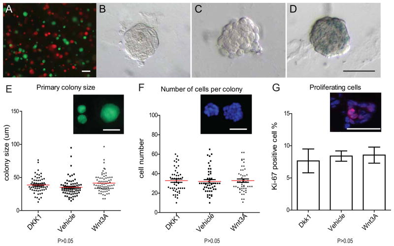
Figure 3. Primary colony formation is not changed by Wnt signaling.
A, FACS-isolated Lin−, CD24+,CD29hi single cells were independently prepared from Actin-GFP mice or Actin-DsRed mice, mixed in a 1:1 ratio and seeded in Matrigel (2x10⁴ cells total in 50 ul pellet). Individual colonies that emerged were exclusively monochromatic, and the number of green and red colonies matched the ratio of the seeded cells (scale bars, 100um). B–D, Lin−, CD24+, CD29hi cells isolated from Axin2lacZ/+ reporter mice were cultured in Matrigel, plus either Dkk1(B), Wnt vehicle control (C) or Wnt3A protein (D). X-gal staining was performed on colonies in 7 days of culture. No Wnt signaling was detectable in Dkk1 or vehicle controls, while the lacZ reporter was activated by Wnt3A protein (F) (scale bar 50um). E, Lin−, CD24+,CD29hi cells isolated from Actin-GFP mice were cultured in Matrigel, plus either Dkk1, Wnt vehicle control or Wnt3A protein. Sizes of the colonies were measured after one week of culture in vitro. In all conditions, there are no differences in the average colony sizes, (which ranged from 20~60um, scale bar, 50um). F, DAPI staining of the colonies in E and cells numbers in each colony were quantified. G, Frozen sections of colonies in E were stained for the proliferation marker Ki-67. There was no difference in the number of Ki-67 positive cells among each condition (P>0.05).

