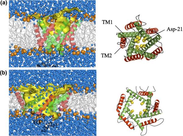
FIGURE 1.
Schematic of numerical simulation of a mechanosensitive ion channel with (a) small and (b) large opening under the action of membrane tension. Left: side view showing the lipid bilayer (white region with red phospholipid head groups) in an aqueous (blue) environment. Right: End view showing the membrane pore. Reproduced with permission from Yefimov et al.93

