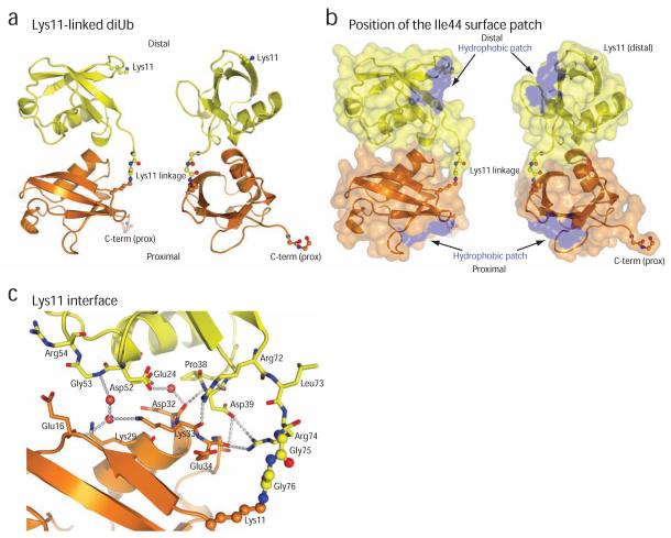
Figure 5.
Crystal structure of Lys11-linked diubiquitin
(a) The crystal structure of Lys11-linked diubiquitin in two orientations. The proximal (orange) and distal (yellow) molecules interact through the ubiquitin helix, and the isopeptide linkage (shown in ball-and-stick representation, with red oxygen and blue nitrogen atoms) is at the surface of the dimer. (b) A semi-transparent surface colored blue for residues Ile44, Leu8 and Val70 shows that the hydrophobic patch is not involved in the interface. (c) Residues at the interface are shown in stick representation, and polar interactions of <3.5Å are shown with dotted lines. Water molecules are shown as purple spheres.

