
Figure 4.
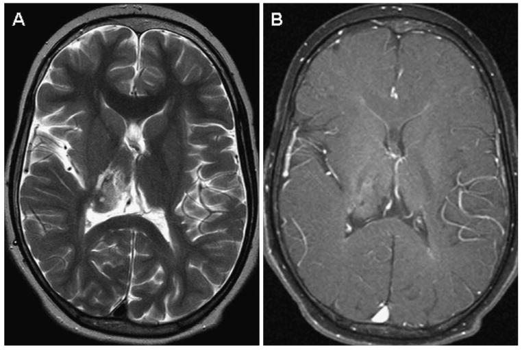
Axial magnetic resonance images of a right thalamic fibrillary astrocytoma status postbiopsy. A, Heterogeneous and hyperintense on T2-weighted images. B, Minimal enhancement on postgadolinium imaging. The patient had left hemiparesis. Because of its location, the tumor was treated with chemotherapy.