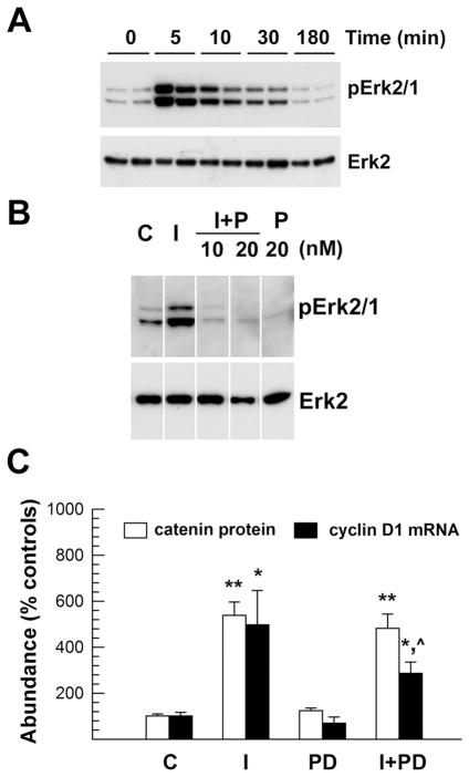
Figure 4.
MAP kinase inhibitor PD98059 suppression of Erk phosphorylation and cyclin D1 mRNA expression stimulated by IGF-I in OL-1 cells. Panel A. Representative Western immunoblot of phosphorylated Erk2/1 (pErk2/1) and total Erk2 in cells treated with 100 ng/ml IGF-I. Treatment duration is indicated at the top of each lane. Panel B. Representative Western immunoblot of phosphorylated Erk2/1 (pErk2/1) and total Erk2 in cells treated with 100 ng/ml IGF-I (I) in the presence or absence of 10 or 20 nM PD98059 (P). Panel C. Quantification of β-catenin protein and cyclin D1 mRNA abundance in cells treated with 100 ng/ml IGF-I and 20 nM PD98059 for 24 hr. Values represent mean ± SE of 4–6 samples. *, P < 0.05, **, P < 0.01, compared to control cells. ^, P < 0.05, compared to cells treated with only IGF-I.

