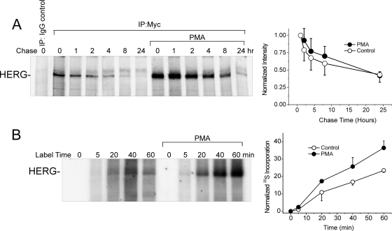
Fig. 7.
PKC effects on synthesis and stability of HERG protein. A, pulse chase of HERG labeled with [35S]cysteine/methionine with and without 10 nM PMA treatment. Graph to the right shows a time-dependent decrease in 35S-HERG normalized to the initial incorporated amount at the beginning of the chase period (n = 4). B, early incorporation of [35S]cysteine/methionine in HERG shown in the first 60 min of labeling with and without PMA treatment. Right, graph represents the densitometry data normalized to total cellular 35S incorporation showing time-dependent new synthesis of HERG (n = 2).

