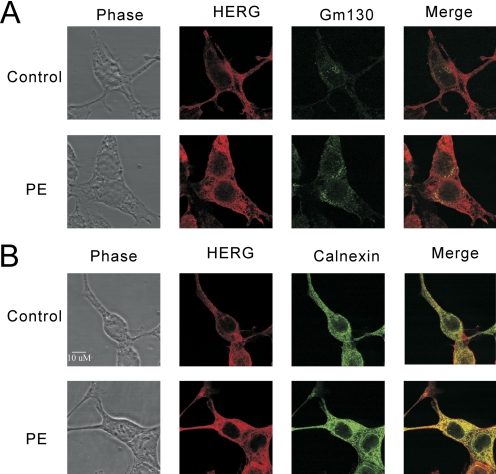
Fig. 8.
Subcellular localization of HERG after 24 h of PKC activation. Confocal immunofluorescence assays with double stating for HERG-myc (red channel) and either the Golgi marker GM130 (A) or the ER marker calnexin (B) (green channel). PMA treatment of 10 nM for 24 h results in globally increased HERG signal in both compartments and on the surface.

