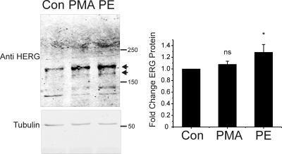
Fig. 9.
α1-AR stimulation enhances ERG channel abundance in rat neonatal cardiac myocytes. Left, anti-HERG immunoblots from isolated rat neonatal cardiac myocytes under control conditions (CON) and after 24 h of treatment with either 10 nM PMA (PMA) or 1 μM PE (PE). ERG channel bands are indicated by arrows. Bottom gel shows tubulin immunoblot from the same gel used to normalize for loading variances. Right, summary data for six experiments with ERG normalized to tubulin densitometry (∗, p < 0.05, n = 6).

