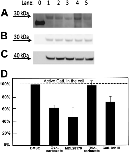
Fig. 5.
Western blot to detect amount of intracellular active Cat-L after treatment with 10 μM inhibitors A, intracellular cysteine protease labeled by DCG-04 was detected at approximately 30 kDa in Western blot. Lane 0, papain (21 kDa), a purified cysteine protease served as a positive control for DCG-04 probe. Lane 1, treated with DMSO only without inhibitor; lane 2, oxocarbazate CID 23631927; lane 3, MDL28170; lane 4, thiocarbazate CID 16725315; lane 5, Cat-L inhibitor III. B, this band was subsequently confirmed to be cathepsin L by anti-cathepsin L rabbit polyclonal antibody. C, protein Loading in each lane was checked using β-actin (molecular mass, 42 kDa) as a control. D, statistical analysis of repeated Western blots was done using Multi Gauge 3.0 (Fujifilm). Data are presented as mean ± S.E. (n = 3).

