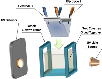
Figure 1.
Schematic drawing of the system used for the measurement of the dielectrophoretic properties of bacterial cells. The UV light (450 nm) was shone between the two slides which were covered with ITO microelectrodes. Any changes in the optical absorbance in a cell suspension by DEP after the generation of nonuniform electric fields at the microelectrodes could be picked up by the UV detector.

