Fig. 1.

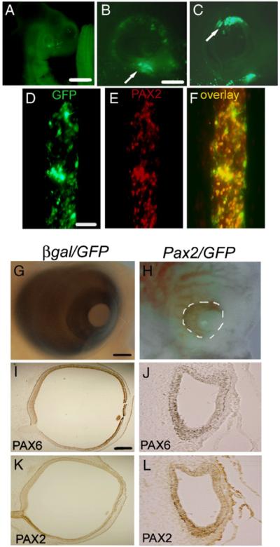
(A–F) Co-expression of GFP with β-gal and Pax2. E3 chick eyes (HH 13–15) were electroporated with β-gal/GFP (10:1) and analyzed at E4. (A, B) GFP is expressed throughout the optic cup, but expression predominates in the ventral portion (arrow). (C) Shows GFP expression in dorsal optic cup (arrow). (D, E) Stage 10 chicks were electroporated with Pax2/GFP (10:1) and sections through forebrain were labeled with GFP (D) and PAX2 (E). (F) Electroporated cells co-express GFP and Pax2. Scale bar (500 μm) in panel A; scale bar (50 μm) in panel B applies to (B, C); scale bar (50 μm) in panel D applies to panels D–F. (G, H) Embryos electroporated at stage 10 were allowed to develop until E6. Embryos electroporated with β-gal/GFP developed normally (G) and showed the typical PAX6 expression patterns throughout the retina (I) and PAX2 expression in the optic nerve (K). In comparison, Pax2/GFP electroporated embryos developed microphthalmia (H; white dashes) in which the optic vesicle-like structure that developed showed very little PAX6 expression (J) and ectopic PAX2 expression (L). Scale bar (50 μm) in panel G applies to panels G, H; scale bar (500 μm) in panel I applies to panels I-L.
