Fig. 2.
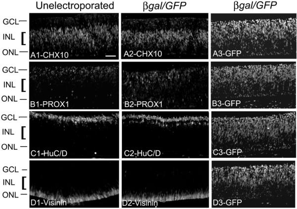
Electroporation has no effect on the normal development of retina. Chick embryos were electroporated with β-gal/GFP at E3 (stage 13–15) and compared with unelectroporated embryos at E8. Immunolabeling was performed on sections through unelectroporated and β-gal/GFP electroporated eyes for CHX10 (Bipolar cells) A1–A2, PROX-1(horizontal cells) B1–B2, HuC/D (amacrine and ganglion cells) C1–C2 and visinin (photoreceptors) D1–D2. A3, B3, C3 and D3 show GFP expression in the respective adjacent sections of the β-gal/GFP electroporated embryos immunolabeled with the retinal markers. ONL; outer nuclear layer, INL; inner nuclear layer, GCL; ganglion cell layer. Scale bar (50 μm) in (A1) applies to (A1–D3).

