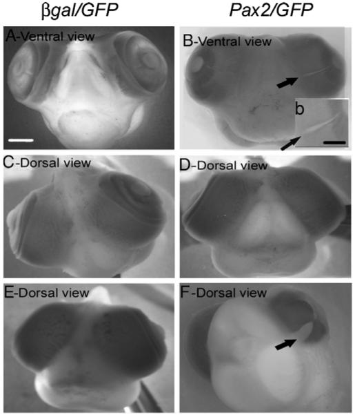
Fig. 3.

Pax2 overexpression leads to lack of choroid fissure closure and ectopic retina. E3 chick embryos were electroporated with β-gal/GFP (A, C, E) or Pax2/GFP (B, D, F) and analyzed at E8. (A) Choroid fissure of β-gal/GFP electroporated embryos showed normal closure at E8, whereas Pax2/GFP electroporated embryos (B) showed the development of a coloboma (B arrow; inset b). panels C and D showed normal development of dorsal eye structures of same embryos shown in panels A and B respectively. Whereas no abnormalities of eye structures were noted in β-gal/GFP electroporated embryos (A, C and E), some Pax2/GFP electroporated embryos also showed regions where RPE appeared to be missing in dorsal eye (F). Scale bar (500 μm) in panel A applies to panels A–F, scale bar (50 μm) in inset (b).
