
Fig. 5.
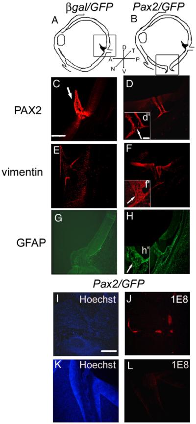
Pax2 expression leads to differentiation of astrocyte processes within the choroid fissure. (A) Shows the schematic of β-gal/GFP electroporated eyes; box represents area shown in panels C, E and G although pecten and optic nerve always do not coincide with each other. (B) shows the schematic of Pax2/GFP electroporated eyes; box represents area shown in panels D, F, H, K and L. (C–H) Pax2 expression leads to differentiation of astrocyte processes within the choroid fissure. PAX2 (C) and vimentin (E) are expressed in the optic nerve, optic nerve head and pecten (arrow in panel C) in control β-gal/GFP electroporated eyes and Pax2/GFP electroporated eyes (not shown). Unlike control E8 eyes, Pax2/GFP electroporated eye showed a persistence of PAX2 (D, d’;arrow) and vimentin (F, f’) expression in cells surrounding the choroid fissure (Hoechst staining of the same region is shown in panel K) and vimentin expression in the processes within the fissure (arrow f’), at E8. Whereas control eyes do not express GFAP in the optic nerve at this stage (G), Pax2/GFP electroporated eye showed GFAP immunolabeled with the cells surrounding the choroid fissure and in the processes of astrocytes within the fissure (H, h’, arrow). Scale bar (50 μm) in panel C applies to panels C–H; scale bar (20 μm) in inset (d’) applies to (d’,f’,h’). (I–L) Cells in the choroid fissure do not express markers characteristic of Schwann cells. Pax2/GFP electroporated embryos with colobomas were immunolabeled for 1E8, a Schwann cell marker. Hoechst label indicates nuclei of cells (I, K). Schwann cells in the peripheral nerves of head can be detected with immunolabel using 1E8 in colobomatous Pax2/GFP electroporated embryos (J), whereas 1E8 antigen cannot be detected in choroid fissure of the same embryo (L). Scale bar (30 μm) in panel I applies to panels I-L.