
Figure 3.
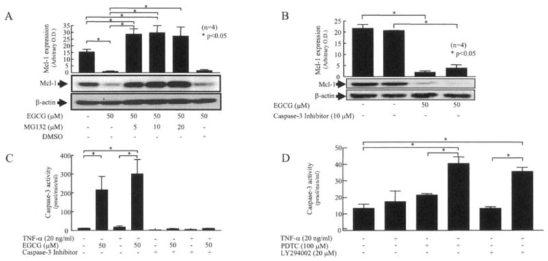
Mechanism of EGCG activation of caspase 3 in RA synovial fibroblasts. A and B, RA synovial fibroblasts (2 × 10⁵/well) were preincubated with MG132 (5–20 μM) or DMSO (A) or with caspase 3 inhibitor (10 μM) (B) for 30 minutes, and then exposed to EGCG (50 μM) for 24 hours. Cells were lysed, and the expression level of Mcl-1 was determined. C, For caspase 3 activity assays, RA synovial fibroblasts (2 × 10⁵/well) were incubated with EGCG (50 μM) alone, TNFα (20 ng/ml) alone, or a combination of EGCG and TNFα for 24 hours in RPMI 1640 plus 1% fetal bovine serum. Cells were lysed in lysis buffer, and 10 μl of lysate was used to estimate caspase 3 activity using commercially available colorimetric assay kits. Results were normalized to protein content in each sample. D, RA synovial fibroblasts were treated with pyrrolidine dithiocarbamate (PDTC; 100 μM) alone, LY294002 (LY; 20 μM) alone, or with a combination of TNFα (20 ng/ml) and PDTC or LY294002 for 24 hours. Cells were lysed with lysis buffer and used to estimate caspase 3 activity. Bars show the mean and SEM from 3 or 4 independent experiments using cells from different donors under similar conditions. * = P < 0.05. OD = optical density (see Figure 1 for other definitions).