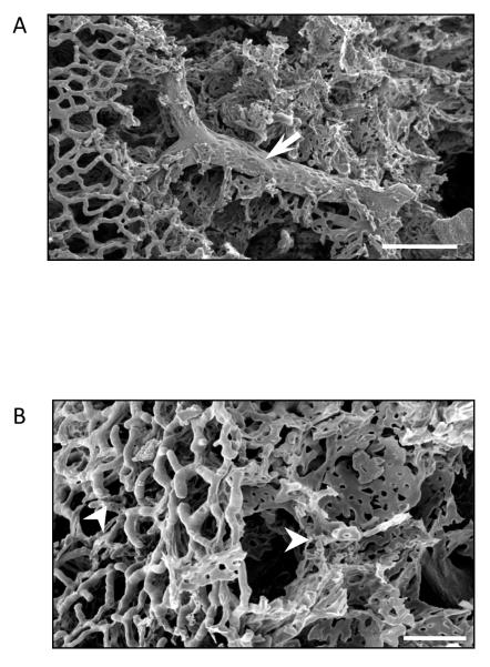
Figure 1.
Scanning electron micrograph of a methylmethacrylate casted lung reveals both the pulmonary macro- and microcirculation. A. Arrow points to extra-alveolar vessel. Scale 100 μm. B. Pulmonary microcirculation. White arrowheads point to arterioles and capillaries. Scale 50 μm. Electron micrographs courtesy of Dr. Diego Alvarez.

