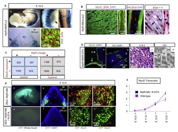
Figure 2. The myf5 Lineage in Myogenesis.
(A) (Aa) The myf5 lineage (blue) in myf5-NN/R-LZ embryos comprises developing embryonic musculature at E10.5, which is predominantly in the (Ab and Ad) myotome but absent in the (Ac and Ad) neural tube. Migratory myogenic populations in the (Ab) limbs (arrow) and (Ad) nonmyogenic dermal precursors (arrows) are shown. (Ae) myf5 expression (red, anti-Myf5) occurs exclusively within the β-gal lineage marker (green, anti-β-gal) in myf5-NN/R-LZ embryos.
(B) The myf5 lineage (red, anti-β-gal) is present within (Ba and Bb) fast-type (green, anti-MyHC) myofibers and (Bc) slow-type (green, anti-MS) myofibers of adult myf5-NN/nLacZ intercostal muscle. (Bd and Be) Enzymatic detection of β-gal activity (red nuclei) reveals a similar distribution of the myf5 lineage, with hematoxylin as counterstain (blue nuclei). Arrows show myonuclei not derived from Myf5.
(C) The distribution of the myf5 lineage (with anti-β-gal) and MyoD expression (using anti-MyoD) was analyzed in epaxial (left, blue box) and hypaxial (right, gray box) regions by cell counting in E12.5 ZEG/myf5-NN embryos. The numbers in boxes are the numbers of nuclei counted. +, presence; −, absence.
(D) (Da) Fast-type (green, anti-MyHC) and (Db) slow-type (green, anti-MS) fibers seen in E18.5 myf5-NN/R-DTA intercostal musculature (D, diaphragm; IC, intercostals; R, ribs) that show normal morphology based on (Dc) H&E staining and (Dd) electron micrograph.
(E) The myf5 lineage (green) demonstrated in (Ea) whole mount and (Eb) sections (green, anti-GFP) in E12.5 ZEG/myf5-NN embryos. (Ec) MyoD (gree,n anti-MyoD) and (Ed) myogenin (green, anti-MyoG) are expressed within and in proximity to the myf5 lineage (red, anti-GFP). Absence of the myf5 lineage in ZEG/myf5-NN/R-DTA embryos is demonstrated in (Ee) whole mount (absence of green signal) and (Ef) sections (absence of green anti-GFP staining). (Eg) MyoD and (Eh) MyoG expression occurs independent of the myf5 lineage in ZEG/myf5-NN/R-DTA.
(F) Semiquantitative RT-PCR for myoD at E10.5, E11.5, E12.5, and E13.5 reveals increased numbers of MyoD transcripts in myf5-NN/R-DTA embryos compared to wild-type by E13.5.

