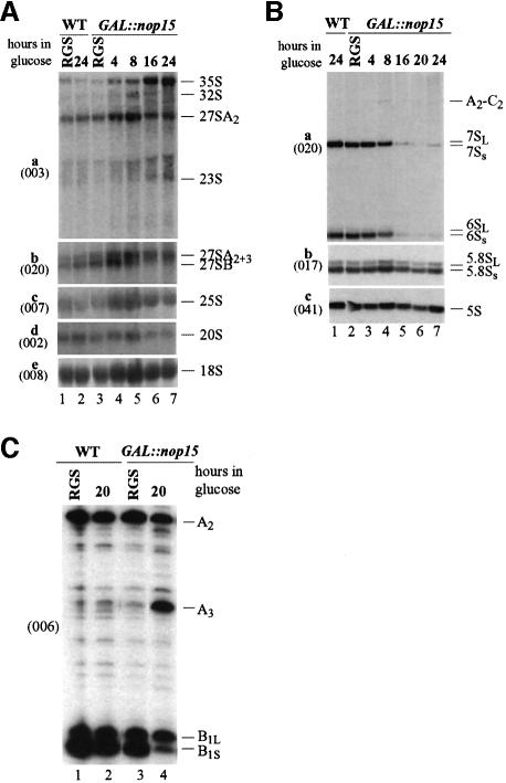
Fig. 3. Analysis of pre-rRNA processing. (A) Northern analysis. Lanes 1 and 2, wild-type strain in RGS medium and 24 h after transfer to glucose; lanes 3–7, GAL::nop15 strain in RGS medium and after transfer to glucose medium for the times indicated. (B) Northern analysis. Lane 1, wild-type strain 24 h after transfer to glucose; lanes 2–7, GAL::nop15 strain in RGS medium and after transfer to glucose medium for the times indicated. RNA was separated on a 1.2% agarose/formaldehyde gel (A) or 8% polyacrylamide/urea gel (B). Probe names are indicated in parentheses on the left. (C) Primer extension using oligo 006, which hybridizes within ITS2, 3′ to site C2 (the 3′ end of the 7S pre-rRNA). Primer extension stops at sites A2, A3, B1S and B1L show levels of the 27SA2, 27SA3, 27SBS and 27SBL pre-rRNAs, respectively. Lanes 1 and 2, wild-type strain in RGS medium and 20 h after transfer to glucose medium; lanes 3 and 4, GAL::nop15 strain in RGS medium and 20 h after transfer to glucose medium.

An official website of the United States government
Here's how you know
Official websites use .gov
A
.gov website belongs to an official
government organization in the United States.
Secure .gov websites use HTTPS
A lock (
) or https:// means you've safely
connected to the .gov website. Share sensitive
information only on official, secure websites.
