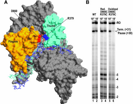
Fig. 6. (A) Structure of the T7RNAP EC (pdb:1msw) with the thumb and fingers subdomains in cyan and orange, respectively. Residues D660 and R379, which form a salt-bridge between the fingers and thumb, are labeled and highlighted in red and blue respectively. (B) Denaturing PAGE of transcription reactions run by forming complexes halted at +14 and then chased with CTP and UTP for 10 s (odd numbered lanes) or 10 min (even numbered lanes) with either wild-type protein (lanes 1 and 2), D660C/R379C double mutant under reducing conditions where the 379–660 disulfide bond is absent (lanes 3 and 4), or the double mutant under conditions which induce formation of the disulfide bond (lanes 5 and 6).

An official website of the United States government
Here's how you know
Official websites use .gov
A
.gov website belongs to an official
government organization in the United States.
Secure .gov websites use HTTPS
A lock (
) or https:// means you've safely
connected to the .gov website. Share sensitive
information only on official, secure websites.
