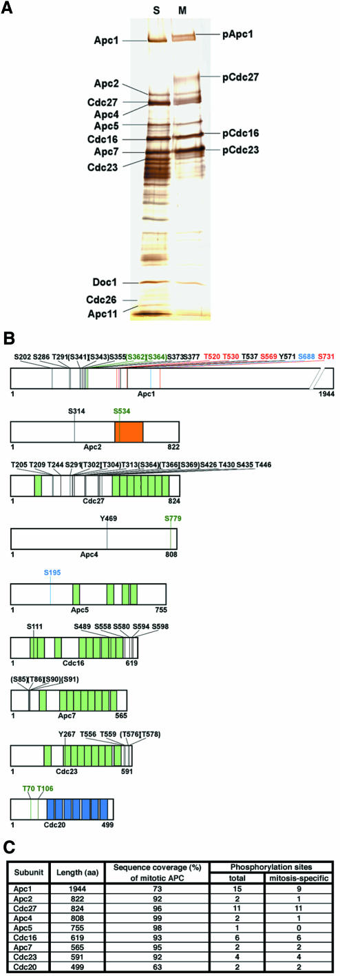
Fig. 1. Phosphorylation sites on human APC. (A) APC was immunoprecipitated from extracts of HeLa cells arrested with nocodazole (M) or hydroxyurea (S), subjected to SDS–PAGE and silver staining. Positions of subunits phosphorylated in mitosis are indicated (pApc1, pCdc27, pCdc16 and pCdc23). (B) Schematic phosphorylation site map of APC subunits derived by nano-HPLC–ESI-MS/MS. Black numbers indicate amino acid residues specifically phosphorylated in mitosis, numbers in parentheses indicate sites that could not be assigned with certainty, blue numbers depict sites found both on mitotic and S-phase APC, red refers to sites only phosphorylated in S-phase. Sites found on mitotic APC that were not covered in the S-phase analysis are shown in dark green. (Light green segments, TPRs; blue segments, WD40 repeats; orange segment, cullin domain.) (C) Summary of phosphorylation sites found on human APC.

An official website of the United States government
Here's how you know
Official websites use .gov
A
.gov website belongs to an official
government organization in the United States.
Secure .gov websites use HTTPS
A lock (
) or https:// means you've safely
connected to the .gov website. Share sensitive
information only on official, secure websites.
