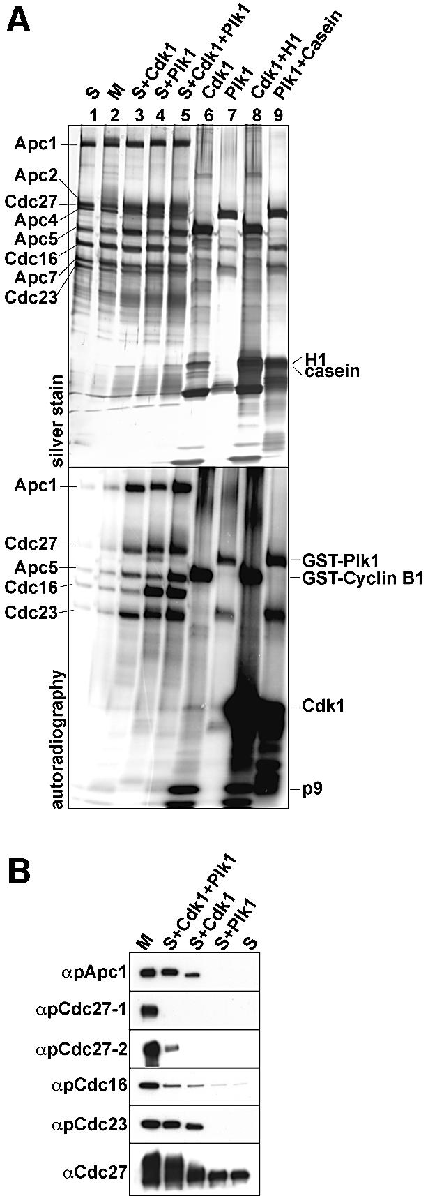
Fig. 3. In vitro phosphorylation of APC by Cdk1 and Plk1. (A) Immunopurified S-phase APC was in vitro phosphorylated using recombinant Cdk1 and Plk1 in the presence of [γ-32P]ATP, as indicated. After stringent washing, the samples were separated by SDS–PAGE, silver stained (upper panel) and analysed by phosphorimaging (lower panel). Lanes 1 and 2, APC incubated in kinase buffer without kinases; lanes 6 and 7, incubation of kinases alone in kinase buffer; lanes 8 and 9, kinases incubated with histone H1 and casein as substrates. (B) APC phosphorylated in vitro as in (A) was analysed by immunoblotting with pApc1-1, pCdc27-1, pCdc27-2, pCdc16 and pCdc23 antibodies. Cdc27 antibodies were used as a control.
